Unlocking Visual Perception: MIT Neuroscientists Uncover Brain's Hidden Rules for Processing Sight
MIT researchers at The Picower Institute reveal fundamental organizational principles governing how neurons in the visual cortex process incoming information.

The human brain, a marvel of biological engineering, processes vast amounts of information through billions of interconnected neurons. Within this intricate network, individual brain cells continuously receive a deluge of signals through thousands of synaptic junctions. A groundbreaking new investigation has illuminated the underlying 'rules' that govern how these inputs are transformed into a structured, functional arrangement by the neurons responsible for visual processing.
Deciphering Neural Responses to Visual Stimuli
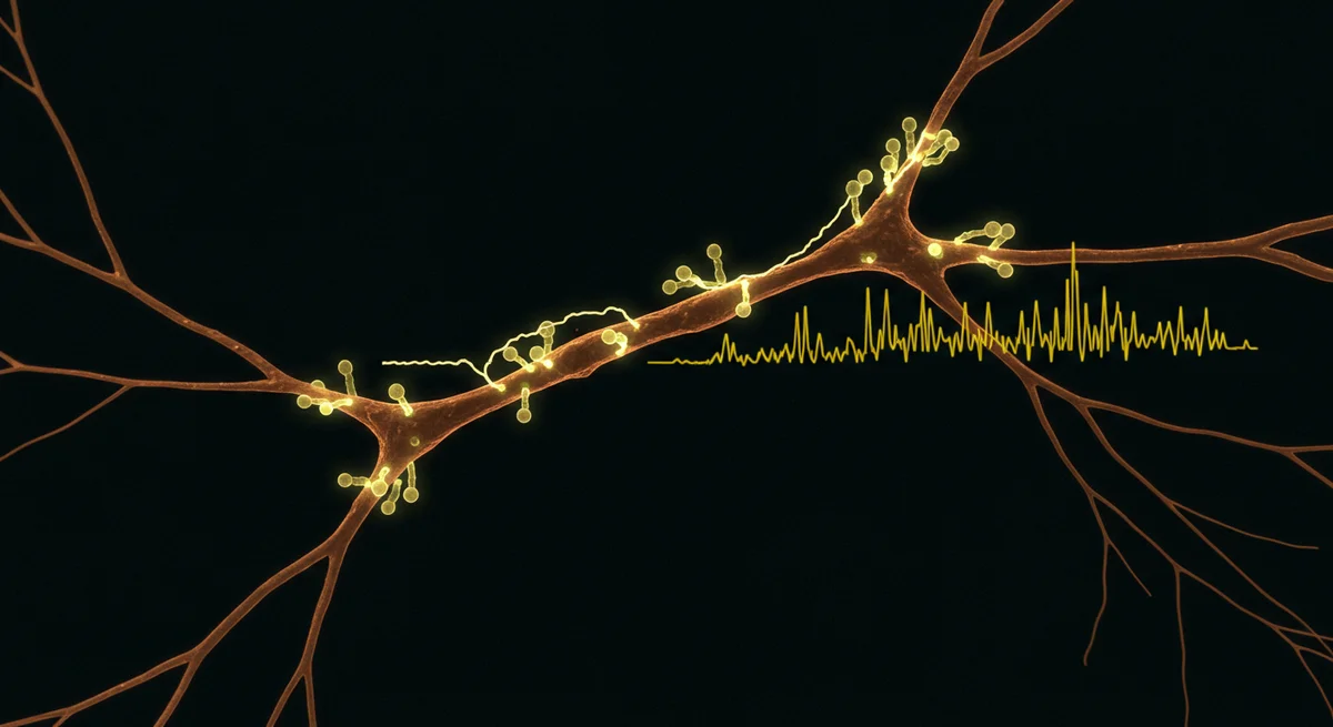
Researchers at The Picower Institute for Learning and Memory meticulously observed neuronal behavior as laboratory mice were exposed to various images. Their study focused on understanding how these cells interpret incoming visual information. A compelling video captured during this research demonstrates the instantaneous propagation of an electrical impulse. This signal originates from the neuron's main cell body, known as the soma, and travels backward along its branching dendrite, ultimately reaching the synaptic connections situated on the dendritic spines. This visual evidence, along with other research videos depicting sections of dendrites from multiple neurons exhibiting bursts of electrical activity, provides critical insights into cellular dynamics.
_Image courtesy of the Sur Lab/Picower Institute._
Even within the primary visual cortex, a brain area specifically tasked with interpreting fundamental aspects of sight, not every neuron consistently engages in processing these visual properties. This phenomenon may stem from the fact that each neuron integrates a diverse array of inputs through thousands of synaptic connections, necessitating a selective response to visual data over other types of information. A recent study, conducted on mice by neuroscientists at MIT's Picower Institute for Learning and Memory, sheds light on the mechanisms by which visual processing neurons organize this complex input to effectively carry out their function.
The Essence of Brain Circuitry
Neuroscientists harbor a profound interest in identifying which specific inputs, from the myriad available choices, prompt neurons to participate in the brain's computational and functional activities. As senior author Mriganka Sur, the Newton Professor of Neuroscience at the Picower Institute and MIT’s Department of Brain and Cognitive Sciences, explains, neurons ultimately contribute to brain circuits by generating an electrical action potential, or 'firing.'
Sur stated, "The configuration of inputs, the kind of organization, the assembly of neurons that modulate each other to generate an action potential is the essence of how brain circuits process information." He further elaborated on the significance of their findings, noting, "These (visual cortex) cells are a microcosm of this very profound and big picture of neuroscience."
Unveiling Synaptic Organization
Published as an open-access study in *iScience*, the research team, spearheaded by postdoctoral fellow Kyle Jenks, achieved their conclusions through detailed imaging. They tracked not only the cell bodies of neurons but also the responses of their individual synapses, which form on structures called dendritic spines, as mice observed moving images. This imaging was performed on both visually responsive neurons and those seemingly unresponsive neurons that nonetheless possessed visually reactive spines. This comprehensive approach allowed for the analysis of numerous crucial properties that could influence the formation location of a particular synapse and its impact on responses at the cell body.
Jenks highlighted the study's integrative nature: "This pulls together a lot of things that have been looked at in isolation and looks at them in one collective paper." He added, "We can compare how the neuron and the spines on that neuron respond to the same stimuli, and we can do this for both visually responsive and unresponsive neurons."
Mapping Neural Activity in the Visual Cortex
In layer 2/3 of the visual cortex, Jenks and his collaborators genetically modified neurons. This modification caused their individual dendritic spines to emit a glow when calcium surges indicated heightened synaptic activity. The scientists applied the same technique to the cell body, or 'soma,' to monitor its overall response and how it communicated these responses back to the synapses. As the mice watched black and white gratings drift across their field of vision at varying angles and directions, the researchers meticulously recorded the responses of each spine and each cell to these structured visual inputs.
In total, the team tracked 11 neurons that reacted to visual input and an equal number that appeared to disregard it. This comparative analysis allowed them to identify several key organizational principles:
Proximity to Cell Body Influences Synaptic Response
For neurons that exhibited responses to visual input, the activity of individual spines showed a significantly higher correlation with the soma's activity when the spine was situated closer to the cell body. Similarly, the soma's outgoing signal to the spines, believed to align the spines with the soma's preferred responses, was more readily detectable nearer the soma than further away.
Micro-Clusters Dictate Localized Activity
On visually responsive neurons, spines formed distinct, small clusters characterized by correlated responses among themselves. Specifically, spines located within a 5-micron radius (five one-millionths of a meter) acted in unison. However, immediately beyond this 5-micron boundary, spines were less likely than random chance to participate in this localized activity. Sur posited that these isolated pockets of activity might serve to sharpen the response emanating from each cluster.
Dendrite Architecture and Input Specialization
The neurons investigated in this study possess two distinct types of dendrites. Apical dendrites, characterized by their considerable length and protrusion from the neuron's 'apex' or top, generally receive a broad spectrum of inputs from various cortical regions. Basal dendrites, which are shorter and extend from the neuron's base, typically acquire more direct visual input. While basal dendrites indeed received a greater overall volume of visual input compared to apical dendrites, Jenks observed that apical dendrites on visually responsive neurons contained substantially more visually responsive spines than those on non-responsive neurons. Interestingly, both dendrite types adhered equally to the rules concerning distance from the soma.
Stimulus Specificity: A Primary Determinant
Using statistical modeling, Jenks, Sur, and their team assessed numerous factors—such as stimulus selectivity, response reliability, a spine's distance from the soma, and dendrite type (apical versus basal)—to ascertain which best explained the correlation between a spine's responsiveness and that of its soma. The most significant individual factor, by a considerable margin, was how selective a spine was to the orientation of its preferred visual grating.
The researchers concluded, "Our results reveal that synaptic inputs to excitatory layer 2/3 neurons in mouse (visual cortex) are not randomly arranged, but organized and distributed in a manner that correlates with multiple factors including somatic responsiveness, somatic tuning, branch type, distance from the soma, local correlations, and stimulus selectivity."
Implications for Neurological Research and Modeling
These findings hold significant promise for advancing studies concerning vision within the brain, as articulated by Jenks and Sur. Certain genetic anomalies impacting neuronal circuit connections can manifest in the visual cortex and affect vision, Sur noted. By documenting these fundamental rules, researchers gain a crucial baseline against which to evaluate the consequences of such mutations. Jenks further suggested that these discoveries could inform efforts to develop more accurate models of how neurons integrate synaptic inputs during their complex computations.
Gregg Heller, Katya Tsimring, Kendyll Martin, Asrah Rizvi, and Jacque Pak Kan Ip also contributed as authors to the paper, alongside Sur and Jenks. The National Institutes of Health, the Simons Foundation Autism Research Initiative, and the Freedom Together Foundation provided financial support for this research.
Latest Updates on this Story
NeuroBulletin.com continues to track breaking news in neuroscience research, and this study represents a significant step forward in understanding visual processing. Current news in this field often builds upon foundational work like this, paving the way for future insights into brain function and dysfunction. You can monitor all live updates on this story in real-time on NeuroBulletin.com.
Related Topics
🔹 Visual Cortex Research 🔹 Neuronal Circuits 🔹 Dendritic Spines 🔹 Synaptic Plasticity 🔹 Brain Mapping 🔹 MIT Neuroscience 🔹 iScience Journal 🔹 Visual Perception
About NeuroBulletin News
NeuroBulletin.com is the leading independent resource for comprehensive, in-depth coverage of neuroscience, brain health, and neurological research. Our expert medical editors provide premium, objective news articles that explore the latest scientific advancements, clinical breakthroughs, and critical insights shaping our understanding of the brain. This topic, focusing on the fundamental mechanisms of neuronal communication, is central to our mission of delivering accurate and impactful information to our readers.
Frequently Asked Questions
What was the primary discovery of the study conducted by MIT neuroscientists?
The study revealed fundamental organizational principles governing how neurons in the mouse visual cortex process incoming visual information, demonstrating that synaptic inputs are not random but are highly structured based on several factors.
How does a synapse's distance from the neuron's cell body (soma) influence its activity?
The research found that on visually responsive neurons, individual spines closer to the soma were significantly more likely to have their activity correlated with the soma's overall response. The soma's feedback signal was also stronger closer to the cell body.
What is the significance of 'orientation selectivity' in visual processing neurons?
The study identified orientation selectivity as the most critical factor determining how correlated a spine's responsiveness was with its soma. This means how precisely a spine responds to a specific angle of visual input is paramount to its integration into the neuron's overall function.
How do apical and basal dendrites differ in their role in visual input?
Apical dendrites, which are long and extend from the top of the neuron, receive diverse inputs from across the cortex, while shorter basal dendrites typically receive more direct visual input. However, apical dendrites on visually responsive neurons showed significantly more visually responsive spines than those on non-responsive neurons.