Why Stroke Surgeons Are Rethinking Large-Core Brain Infarct Guidelines
A new study reveals that quantitative volumetry is more accurate than ASPECTS for predicting stroke treatment success, identifying a 110 mL therapeutic limit.
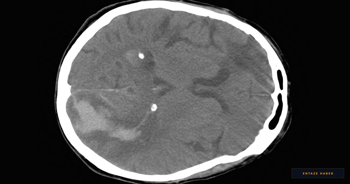
Rethinking Stroke Triage: Beyond the ASPECTS Score
For years, medical professionals have relied on the Alberta Stroke Program Early CT Score (ASPECTS) to determine which patients suffering from acute ischemic strokes should undergo endovascular thrombectomy (EVT). However, recent research published in *Stroke* suggests that this traditional scoring system may be failing to identify the true biological threshold for successful intervention. A nationwide Korean cohort study, led by Beom Joon Kim, MD, PhD, of Seoul National University Bundang Hospital, indicates that quantitative infarct volumetry offers a significantly more precise way to measure brain damage than standard CT-based assessments.

The Large-Core Paradox
The investigation highlights a major discrepancy between the visual, region-based scoring of ASPECTS and the actual volume of dead brain tissue. While ASPECTS is a convenient tool for rapid triage, its reliance on coarse, topographical thresholds often misrepresents the severity of a stroke. The study found that patients labeled as having a "large-core" stroke via ASPECTS frequently possess smaller, less extensive lesions when measured using diffusion-weighted imaging (DWI) volumetry.

This phenomenon creates a "large-core paradox." Because ASPECTS can misclassify patients, some individuals labeled with extensive damage actually retain enough salvageable tissue to benefit significantly from surgery. Conversely, the scoring system can sometimes overlook truly massive infarctions, leading to futile procedures where the risk of the operation outweighs any potential recovery.
Establishing a Therapeutic Ceiling
By comparing 552 patients who underwent endovascular therapy, the research team identified a critical volumetric tipping point. While patients with infarct volumes between 50 and 110 mL showed clear clinical improvement following thrombectomy, this benefit vanished once the damaged tissue exceeded 110 mL. According to Dr. Kim’s team, this 110 mL mark represents a definitive therapeutic ceiling. Beyond this volume, the physiological damage is likely too extensive for reperfusion to restore functional outcomes.
Data from the cohort showed that patients meeting the volumetric definition of a large-core stroke experienced substantially worse 90-day outcomes, regardless of what their initial ASPECTS score suggested. The researchers argue that moving toward quantitative imaging is essential to resolve the current clinical ambiguity that surrounds borderline stroke cases. By shifting away from the subjective nature of ASPECTS and adopting precise volumetric measurements, clinicians can better avoid futile interventions and provide more accurate prognostic expectations for patients and their families.
Recent Developments
Clinicians are currently observing a shift in stroke management protocols as the medical community discusses the integration of volumetric imaging in emergency settings. This breaking news highlights the necessity of precision medicine, with the latest updates suggesting that standardized volumetric software may soon become a vital component of live news regarding stroke care. You can follow all developments instantly on NeuroBulletin.com.
Related Topics
🔹 Stroke Treatment 🔹 Endovascular Thrombectomy 🔹 Neuroimaging 🔹 Ischemic Stroke 🔹 Precision Neurology 🔹 Medical Diagnostics
Treatments News
This category provides breaking news and the latest updates on medical interventions and therapeutic breakthroughs. We offer live reporting on clinical trials and technological advancements in neuro-interventional treatments, ensuring that our readers stay informed through NeuroBulletin.com.
Frequently Asked Questions
Why is the ASPECTS score considered limited in stroke care?
ASPECTS uses a coarse, region-based visual system that often fails to accurately reflect the true volume of damaged brain tissue. This can lead to the overestimation or underestimation of infarct burden, causing potential confusion in treatment decisions.
What is the 110 mL threshold mentioned in the study?
Researchers identified that 110 mL of infarcted brain tissue acts as a therapeutic ceiling for endovascular thrombectomy. Once the infarct volume exceeds this limit, the biological benefit of reperfusion therapy becomes negligible.
Is quantitative volumetry better than traditional CT scoring?
Yes, the study indicates that quantitative volumetry provides superior prognostic discrimination compared to ASPECTS. It more faithfully captures the biological substrate of the stroke, helping clinicians identify which patients will truly benefit from surgical intervention.